


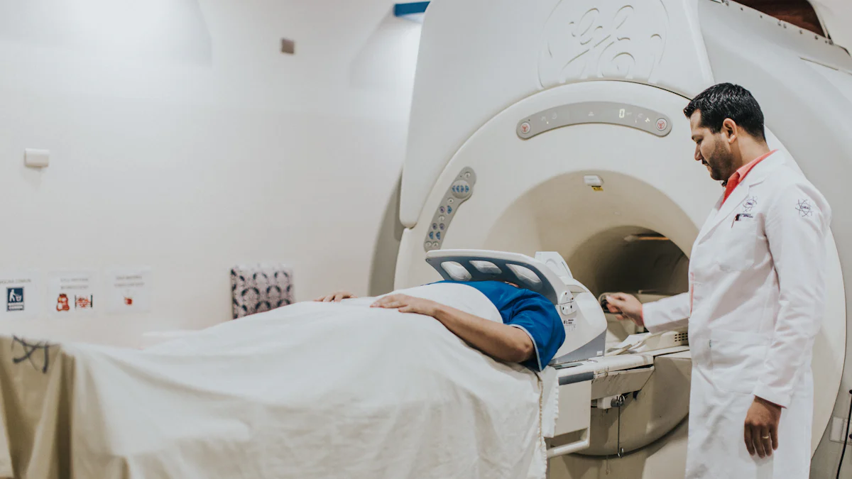
Electron beam angiography represents a groundbreaking advancement in medical imaging. This technique utilizes electron beams to produce detailed images of blood vessels, offering a non-invasive approach to diagnosing various conditions. Primarily, it excels in the realm of cardiovascular health, providing precise insights into coronary artery disease. Unlike traditional methods, electron beam angiography delivers rapid and accurate results, enhancing the diagnostic process. Its ability to measure coronary calcification makes it an invaluable tool in assessing heart health. As a result, electron beam angiography has become a cornerstone in modern diagnostics, revolutionizing how medical professionals approach patient care.
Key Takeaways
- Electron beam angiography is a non-invasive imaging technique that provides detailed images of blood vessels, particularly useful in diagnosing coronary artery disease.
- This technology offers rapid imaging capabilities, allowing for quick diagnosis and timely intervention, which can significantly improve patient outcomes.
- With high-resolution imaging, electron beam angiography enhances diagnostic accuracy, instilling confidence in clinicians when assessing cardiovascular and neurological conditions.
- The technique reduces patient discomfort and risk by eliminating the need for catheterization, making it a safer alternative to traditional invasive methods.
- Ongoing advancements in technology, including AI integration, promise to further enhance the accuracy and efficiency of electron beam angiography in medical diagnostics.
- Despite its advantages, the high cost and specialized training required for operation can limit accessibility in some medical facilities.
- As research expands, electron beam angiography may find new applications in fields like oncology and personalized medicine, broadening its impact on patient care.
Understanding Electron Beam Angiography
The Technology Behind Electron Beam Angiography
How Does Electron Beams Work?
Electron-beam computed tomography (EBCT) represents a significant leap in medical imaging technology. This technique employs a focused beam of electrons to scan the body, creating detailed images of internal structures. Unlike traditional X-ray methods, EBCT does not require mechanical movement of the scanner, which allows for rapid image acquisition. The electron beam generates X-rays that pass through the body, and detectors capture the resulting data to form high-resolution images. This process enables clinicians to visualize blood vessels and other tissues with remarkable clarity.
Imaging Process and Techniques
The imaging process in electron beam CT involves several steps. Initially, the electron beam targets a tungsten target, producing X-rays. These X-rays penetrate the body and are absorbed by detectors positioned around the patient. The data collected by these detectors is then processed to create cross-sectional images of the body. EBCT’s ability to capture images quickly makes it particularly useful for cardiac imaging, where motion can blur images. Studies have shown that EBCT provides high spatial resolution and electrocardiographic-triggered images, which are crucial for accurate coronary assessments.
Historical Development and Adoption
Evolution of Angiography Techniques
Angiography has evolved significantly over the years. Initially, invasive techniques dominated the field, requiring catheter insertion into blood vessels. These methods, while effective, posed risks and discomfort to patients. The advent of electron beam CT marked a turning point, offering a non-invasive alternative. This innovation reduced the need for catheterization, minimizing patient risk and discomfort. As technology advanced, EBCT became a reliable tool for evaluating aortic disease and pulmonary vessels, demonstrating excellent resolution along the z-axis.
Milestones in Electron Beam Angiography
Several milestones highlight the development of electron beam angiography. Early studies demonstrated the feasibility of using EBCT for coronary angiography, comparing its results with traditional invasive methods. Although initial images were suboptimal for clinical application, ongoing research explored different aspects and limitations of EBCT. Over time, improvements in imaging techniques and technology enhanced the quality and reliability of EBCT images. Today, electron beam CT serves as a valuable tool in diagnosing coronary artery disease, offering a non-invasive, accurate, and efficient alternative to traditional methods.
Applications in Diagnostics
Electron beam angiography has revolutionized the field of medical diagnostics, offering unparalleled insights into various diseases. Its applications span across multiple domains, providing clinicians with powerful tools for accurate diagnosis and treatment planning.
Cardiovascular Diagnostics
Coronary Artery Disease Detection
Electron beam angiography excels in detecting coronary artery disease. This non-invasive diagnostic test provides detailed images of the coronary arteries, allowing clinicians to identify blockages and assess the severity of the disease. The technique’s high sensitivity and specificity make it a preferred choice for evaluating coronary artery conditions. By measuring coronary artery calcium, electron beam angiography offers a reliable method for assessing the risk of obstructive coronary artery disease. This capability enhances the diagnostic process, enabling timely intervention and improved patient outcomes.
Aneurysm Identification
In addition to coronary artery disease detection, electron beam angiography plays a crucial role in identifying aneurysms. The technique’s rapid imaging capabilities and high-resolution images allow for precise visualization of vascular structures. Clinicians can detect aneurysms in the coronary arteries with greater accuracy, facilitating early diagnosis and intervention. This application underscores the importance of electron beam angiography in cardiovascular diagnostics, providing a comprehensive view of the heart’s vascular system.
Neurological Applications
Stroke Diagnosis
Electron beam angiography extends its diagnostic prowess to neurological applications, particularly in stroke diagnosis. The technique’s ability to capture detailed images of cerebral blood vessels aids in identifying blockages or abnormalities that may lead to a stroke. Its high sensitivity and specificity ensure accurate diagnosis, enabling clinicians to devise effective treatment plans. By offering a non-invasive alternative to traditional methods, electron beam angiography enhances the diagnostic process, reducing the time to treatment and improving patient outcomes.
Brain Aneurysm Detection
Detecting brain aneurysms is another critical application of electron beam angiography. The technique provides clear images of the brain’s vascular structures, allowing clinicians to identify aneurysms with precision. This capability is vital for early diagnosis and intervention, preventing potential complications. Electron beam angiography’s high sensitivity and specificity make it an invaluable tool in neurological diagnostics, offering a safe and efficient method for assessing brain health.
Other Medical Applications
Peripheral Vascular Disease
Beyond cardiovascular and neurological applications, electron beam angiography proves beneficial in diagnosing peripheral vascular disease. The technique’s ability to visualize blood vessels in the extremities aids in identifying blockages or abnormalities. Clinicians can assess the severity of the disease and plan appropriate interventions, improving patient outcomes. This application highlights the versatility of electron beam angiography in medical diagnostics, providing comprehensive insights into vascular health.
Pulmonary Embolism
Electron beam angiography also plays a significant role in diagnosing pulmonary embolism. The technique’s rapid imaging capabilities and high contrast enhancement allow for detailed visualization of pulmonary vessels. Clinicians can identify emboli with high sensitivity and specificity, ensuring accurate diagnosis and timely treatment. This application underscores the importance of electron beam angiography in respiratory diagnostics, offering a reliable method for assessing pulmonary health.
Advantages Over Traditional Methods
Electron beam angiography offers several advantages over traditional methods, making it a preferred choice in modern diagnostics. Its unique capabilities enhance the diagnostic process, providing clinicians with valuable insights into various medical conditions.
Speed and Efficiency
Rapid Imaging Capabilities
Electron beam angiography excels in speed, allowing for rapid imaging of the heart and blood vessels. The technology’s ability to capture high-resolution images quickly is particularly beneficial in cardiac diagnostics. Clinicians can obtain detailed images of the coronary arteries without the need for prolonged procedures. This efficiency reduces the time required for diagnosis, enabling timely intervention and improving patient outcomes.
Reduced Procedure Time
The noninvasive nature of electron beam angiography significantly reduces procedure time compared to traditional methods. Patients benefit from shorter appointments, minimizing discomfort and anxiety. The quick imaging process also allows healthcare providers to accommodate more patients, enhancing the overall efficiency of medical facilities. This advantage makes electron beam angiography an attractive option for both patients and clinicians.
Accuracy and Precision
High-Resolution Imaging
Electron beam angiography provides high-resolution images that enhance diagnostic accuracy. The technology’s ability to capture detailed images of the coronary arteries allows clinicians to identify blockages and assess the severity of coronary artery disease with precision. This level of detail is crucial for accurate diagnosis and effective treatment planning. By offering superior image quality, electron beam angiography improves the diagnostic accuracy of coronary artery disease assessments.
Enhanced Diagnostic Confidence
The precision of electron beam angiography instills confidence in clinicians when diagnosing coronary artery disease. The technique’s high sensitivity and specificity ensure that diagnoses are accurate and reliable. Clinicians can make informed decisions based on clear and detailed images, reducing the likelihood of misdiagnosis. This enhanced diagnostic confidence leads to better patient care and improved outcomes.
Safety and Patient Comfort
Non-Invasive Nature
Electron beam angiography is a noninvasive testing method, offering a safer alternative to traditional invasive procedures. Patients do not require catheter insertion, reducing the risk of complications and discomfort. This noninvasive assessment approach makes the diagnostic process more comfortable for patients, encouraging them to undergo necessary evaluations without hesitation.
Lower Radiation Exposure
Compared to other imaging techniques, electron beam angiography exposes patients to lower levels of radiation. This reduction in radiation exposure enhances patient safety, particularly for those requiring frequent diagnostic tests. The technique’s ability to provide accurate results with minimal radiation makes it a preferred choice for long-term monitoring of coronary artery disease.
Limitations and Considerations
Electron beam angiography, while revolutionary, presents certain limitations and considerations that medical professionals must acknowledge. Understanding these factors ensures informed decision-making in clinical settings.
Cost and Accessibility
Equipment and Maintenance Costs
The high cost of electron beam angiography equipment poses a significant barrier to widespread adoption. Hospitals and clinics must invest in advanced machinery, which requires substantial financial resources. Additionally, maintaining this sophisticated technology demands regular servicing and skilled technicians, further increasing operational expenses. These costs can limit the availability of electron beam angiography, particularly in resource-constrained settings.
Availability in Medical Facilities
Not all medical facilities can offer electron beam angiography due to its specialized nature. Larger hospitals and research centers are more likely to have the necessary infrastructure. Smaller clinics may lack access, restricting patient options for non-invasive coronary diagnostics. This disparity in availability highlights the need for strategic planning to ensure broader access to this valuable diagnostic tool.
Technical Limitations
Image Artifacts
Electron beam angiography, despite its advanced capabilities, can produce image artifacts. These artifacts may arise from partial volume effects, especially when imaging very small vessel diameters. Such limitations can affect the accuracy of the diagnosis, as evidenced by studies comparing vessel diameters in electron beam tomography and quantitative coronary angiography. Clinicians must interpret results with caution, considering potential discrepancies in image quality.
Operator Expertise Required
Operating electron beam angiography equipment requires specialized training and expertise. Technicians and radiologists must possess a deep understanding of the technology to ensure accurate image acquisition and interpretation. The need for skilled operators can limit the use of this diagnostic method in facilities lacking adequately trained personnel. Continuous education and training programs are essential to address this challenge.
Patient-Specific Considerations
Suitability for Different Patient Groups
Electron beam angiography may not be suitable for all patient groups. Factors such as age, health conditions, and specific medical histories can influence the appropriateness of this diagnostic approach. For instance, patients with high calcium scores may benefit from preventive interventions, but routine use of electron beam computed tomography remains unjustified for individual patients without clear additional value. Clinicians must evaluate each case individually to determine the best diagnostic strategy.
Contraindications and Risks
Certain contraindications and risks accompany the use of electron beam angiography. Patients with specific medical conditions or allergies to contrast agents may face complications. Additionally, while the radiation exposure is lower than traditional methods, it still poses a risk, particularly for patients requiring frequent imaging. Understanding these risks is crucial for ensuring patient safety and optimizing diagnostic outcomes.
Future Prospects and Innovations
Technological Advancements
Integration with AI and Machine Learning
Electron beam angiography stands on the brink of a technological revolution. The integration of artificial intelligence (AI) and machine learning promises to enhance diagnostic accuracy and efficiency. AI algorithms can analyze vast amounts of imaging data, identifying patterns and anomalies that might escape human detection. This capability allows clinicians to make more informed decisions, improving patient outcomes. Machine learning models can also predict disease progression, offering personalized treatment plans. These advancements position electron beam angiography as a cutting-edge tool in modern diagnostics.
Improvements in Imaging Techniques
Continuous improvements in imaging techniques drive the evolution of electron beam angiography. Researchers focus on enhancing image quality and reducing artifacts, ensuring clearer and more precise visualizations. Innovations in detector technology and image processing algorithms contribute to these improvements. As a result, clinicians can obtain high-resolution images with greater detail, facilitating accurate diagnoses. These advancements not only improve the reliability of electron beam angiography but also expand its potential applications in various medical fields.
Expanding Clinical Applications
Emerging Areas of Research
The scope of electron beam angiography extends beyond traditional applications. Researchers explore its potential in new areas, such as oncology and orthopedics. In oncology, electron beam angiography could aid in tumor detection and monitoring, providing detailed images of vascular structures within tumors. This application could enhance cancer diagnosis and treatment planning. In orthopedics, the technique might assist in assessing bone vascularization, offering insights into conditions like osteoporosis. These emerging areas of research highlight the versatility and adaptability of electron beam angiography.
Potential for Personalized Medicine
Electron beam angiography holds promise for advancing personalized medicine. By providing detailed insights into individual vascular health, the technique enables tailored treatment plans. Clinicians can assess a patient’s unique risk factors and disease progression, customizing interventions accordingly. This personalized approach enhances treatment efficacy and patient satisfaction. As electron beam angiography continues to evolve, its role in personalized medicine will likely expand, offering new possibilities for individualized care.
Conclusion
Electron beam angiography stands as a transformative force in medical diagnostics. This technique offers unparalleled insights into cardiovascular and neurological health. Its non-invasive nature and rapid imaging capabilities make it a preferred choice for clinicians. Electron beam computed tomography (EBCT) provides high-resolution images, enhancing diagnostic accuracy and confidence. Studies highlight its potential as a screening tool for coronary artery disease. As technology advances, electron beam angiography will continue to revolutionize patient care, offering precise and efficient diagnostic solutions. Its power and potential in modern diagnostics remain undeniable.