


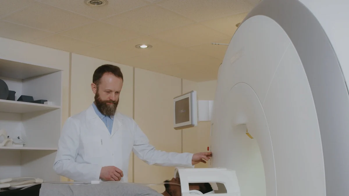
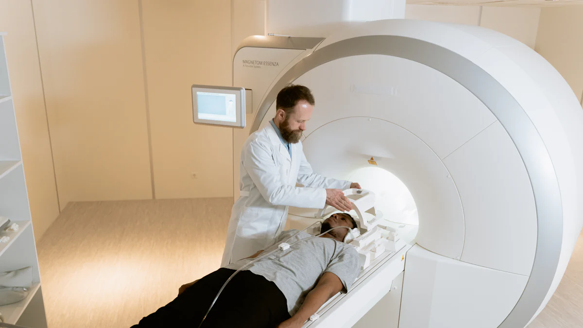
Medical imaging plays a crucial role in diagnosing and monitoring various health conditions. Electron beam CT scan and traditional medical CT represent two advanced technologies with distinct features. Electron beam CT scan excels in speed and precision, capturing up to 20 images per second, making it ideal for cardiac imaging. Traditional medical CT, on the other hand, offers superior spatial resolution and versatility across a wide range of diagnostic applications. Understanding these differences helps patients and healthcare providers make informed decisions tailored to specific medical needs.
Key Takeaways
- Electron Beam CT scan captures images at an impressive rate of up to 20 images per second, making them ideal for cardiac imaging and minimizing motion artifacts.
- Traditional CT scan offers exceptional spatial resolution and versatility, making them suitable for diagnosing a wide range of conditions across various body parts.
- When choosing between imaging technologies, consider factors like image quality, safety, and cost to ensure the best option for your medical needs.
- Electron Beam CT is particularly beneficial for patients requiring frequent imaging due to its lower radiation exposure compared to some traditional methods.
- Both imaging technologies play vital roles in diagnostics; Electron Beam CT excels in specialized fields like cardiology, while Traditional CT remains the gold standard for general medical imaging.
- Advancements in cone beam CT technology have improved traditional CT’s efficiency, making it a practical choice for dental and maxillofacial imaging.
- Consulting healthcare professionals is essential to determine the most suitable imaging option based on individual medical requirements and desired clinical outcomes.
Overview of Electron Beam CT and Traditional Medical CT
What is an Electron Beam CT Scan?
An electron beam CT scan represents a significant advancement in medical imaging technology. Unlike traditional methods, this type of computed tomography uses an electron beam to generate high-resolution images. The electron beam targets a stationary ring around the patient, eliminating the need for mechanical movement. This setup reduces motion artifacts and enhances image clarity.
One of the standout features of this technology is its speed. The machine can capture up to 20 images per second, making it ideal for imaging moving organs like the heart. This rapid image acquisition minimizes distortion caused by cardiac motion. Additionally, its high temporal resolution, which can reach up to 8000 frames per second, allows for precise visualization of dynamic processes within the body.
The electron beam CT scan is particularly effective in detecting and quantifying coronary artery calcification. This capability makes it a valuable tool for diagnosing conditions such as atherosclerosis and coronary artery disease. Its ability to provide detailed images with minimal operator intervention ensures consistent results, enhancing its reliability in clinical settings.
What is a Traditional Medical CT?
A traditional medical CT scan, also known as a conventional CT, has been a cornerstone of diagnostic imaging for decades. It employs a fan-shaped X-ray beam that rotates around the patient to capture multiple image slices. These slices are then reconstructed into detailed cross-sectional images of the body.
Traditional CT scanners excel in versatility. They can image a wide range of body parts, from the brain to the abdomen, with exceptional spatial resolution. This adaptability makes them suitable for diagnosing various conditions, including fractures, tumors, and internal bleeding. The technology’s ability to produce high-quality images ensures accurate diagnoses across multiple medical disciplines.
However, the imaging process in traditional CT scanners involves mechanical rotation, which may result in motion artifacts when imaging moving organs. Despite this limitation, advancements in cone beam CT technology have improved the speed and precision of these systems. Cone beam CT scanners, for instance, use a cone-shaped X-ray beam to capture volumetric data in a single rotation, reducing scan times and improving efficiency.
Key Technical Differences
Imaging Technology and Mechanism
The imaging technology behind electron beam CT scan and traditional medical CT differs significantly. Electron beam CT employs an electron beam directed at a stationary tungsten target, which eliminates the need for mechanical rotation. This design reduces motion artifacts and enhances the clarity of images, particularly for moving organs like the heart. In contrast, traditional CT scanners rely on a rotating X-ray tube that moves around the patient to capture multiple image slices. These slices are then reconstructed into detailed cross-sectional images using advanced algorithms.
Cone beam CT scanners, a variation of traditional CT, use a cone-shaped X-ray beam to capture volumetric data in a single rotation. This approach improves efficiency and reduces scan times. However, conventional CT remains the gold standard for general medical imaging due to its ability to provide high image quality and superior contrast resolution. The choice of technology often depends on the specific clinical requirements and the anatomical region being examined.
Speed and Precision in Imaging
Speed and precision are critical factors in medical imaging. The electron beam CT scan excels in this area, offering fast, accurate imaging with subsecond scanning times. It can capture up to 20 images per second, making it ideal for cardiac imaging and other applications requiring high temporal resolution. This capability ensures that dynamic processes, such as blood flow or organ movement, are captured without distortion.
Traditional CT scanner, while slightly slower, has also seen advancements in speed. Modern designs, including cone beam CT, can acquire multiple slices in a single rotation, reducing overall scan times. Despite these improvements, traditional CT may still face challenges when imaging rapidly moving tissues, such as the heart. However, its optimized contrast resolution and ability to visualize fine anatomical details make it indispensable for a wide range of diagnostics.
Suitability for Specific Medical Applications
The suitability of each technology depends on the medical application. The electron beam CT scan is particularly effective for cardiac imaging, where speed and precision are paramount. Its ability to quantify coronary artery calcification makes it a valuable tool for diagnosing coronary artery disease and assessing cardiovascular risk. Additionally, its lower radiation exposure compared to some traditional methods enhances patient safety, especially for repeated scans.
Traditional CT scanner offers unmatched versatility. They are widely used for imaging various body parts, including the brain, abdomen, and musculoskeletal system. Their high spatial resolution and superior image quality make them ideal for detecting fractures, tumors, and internal bleeding. Cone beam CT, a subset of traditional CT, is often preferred in dental and maxillofacial imaging due to its ability to provide detailed three-dimensional views of bone and soft tissue structures.
In clinical practice, the choice between these technologies often depends on the desired clinical outcomes. For example, conventional CT is recommended for general medicine and surgery due to its broad applicability and high-quality imaging. On the other hand, electron beam CT is favored in specialized fields like cardiology, where rapid imaging and minimal motion artifacts are crucial.
Benefits and Applications of Each Technology
Benefits of Electron Beam CT Scan
The electron beam CT scan offers several advantages that make it a valuable tool in modern medical imaging. Its exceptional speed stands out as a defining feature. By capturing up to 20 images per second, this technology ensures precise imaging of moving organs, such as the heart. This capability minimizes motion artifacts, which often compromise the clarity of images in other imaging methods. The high temporal resolution further enhances its ability to visualize dynamic processes, such as blood flow, with remarkable accuracy.
Another significant benefit lies in its reduced radiation exposure compared to some traditional methods. This makes the electron beam CT scan a safer option for patients requiring repeated imaging, such as those undergoing cardiac monitoring. Additionally, its stationary design eliminates the need for mechanical rotation, ensuring consistent image quality and reliability. This feature proves particularly useful in clinical settings where precision is critical.
The electron beam CT scan excels in cardiac diagnostics. It effectively detects and quantifies coronary artery calcification, aiding in the early diagnosis of coronary artery disease. This capability allows healthcare providers to assess cardiovascular risk and implement timely interventions. Its ability to deliver high-quality images with minimal operator intervention ensures consistent and accurate results, making it a preferred choice in specialized fields like cardiology.
Benefits of Traditional Medical CT
The traditional CT scanner remains a cornerstone of diagnostic imaging due to its versatility and adaptability. It provides exceptional spatial resolution, enabling detailed visualization of various anatomical structures. This high resolution ensures accurate identification of abnormalities, such as fractures, tumors, and internal bleeding. Its ability to produce high-quality images across multiple body regions makes it indispensable in general diagnostics.
One of the key strengths of the traditional CT scanner lies in its broad applicability. It can image a wide range of tissues, from soft tissue structures to dense bones, with remarkable contrast resolution. This adaptability allows healthcare providers to diagnose conditions affecting different parts of the body, including the brain, abdomen, and musculoskeletal system. For instance, in stroke diagnosis, the traditional CT scanner effectively identifies hemorrhagic strokes, guiding timely medical interventions.
Advancements in cone beam CT technology have further enhanced the capabilities of traditional CT scanners. Cone beam CT scanners use a cone-shaped X-ray beam to capture volumetric data in a single rotation, reducing scan times and improving efficiency. This innovation has expanded the applications of traditional CT, particularly in dental and maxillofacial imaging, where detailed three-dimensional views of bone and tissue structures are essential.
Common Applications and Use Cases
Both electron beam CT and traditional CT scanners play vital roles in medical diagnostics, each excelling in specific applications. The electron beam CT scan is particularly effective in cardiac imaging. Its speed and precision make it ideal for assessing coronary artery disease and quantifying coronary artery calcification. This technology also proves valuable in emergency settings, where rapid imaging is crucial for timely decision-making.
Traditional CT scanners, on the other hand, offer unmatched versatility. They are widely used in general medicine and surgery for diagnosing a variety of conditions. For example, in abdominal imaging, traditional CT effectively detects appendicitis in patients presenting with acute abdominal pain. Its ability to provide high-quality images ensures accurate diagnoses, even in complex cases.
Cone beam CT technology has further expanded the scope of traditional CT applications. Cone beam CT scanners are commonly used in dental and maxillofacial imaging, providing detailed three-dimensional views of bone and tissue structures. This capability proves invaluable in planning surgical procedures and assessing dental conditions.
In clinical practice, the choice between these technologies often depends on the desired clinical outcomes. Electron beam CT is favored in specialized fields like cardiology, where speed and precision are paramount. Traditional CT scanners, with their superior contrast resolution and broad applicability, remain the gold standard for general diagnostics. Both technologies contribute significantly to improving patient care and advancing medical imaging.
Safety and Radiation Exposure Comparison
Radiation Levels and Patient Safety
Radiation exposure remains a critical factor when evaluating the safety of imaging technologies. Electron beam CT (EBCT) offers a distinct advantage by utilizing lower doses of radiation compared to some traditional methods. This feature makes it a safer option for patients requiring frequent imaging, such as those undergoing cardiac monitoring. The stationary design of EBCT eliminates the need for mechanical rotation, which contributes to its ability to deliver high-quality images while minimizing radiation levels.
Conventional CT, while versatile and widely used, often involves higher radiation doses due to its reliance on a rotating X-ray tube. This design allows for detailed cross-sectional images of anatomical structures but may expose patients to increased radiation levels, especially during repeated scans. However, advancements in technology, such as cone beam CT, have helped reduce radiation exposure in certain applications. These improvements enhance patient safety without compromising image resolution or diagnostic accuracy.
Healthcare providers must weigh the benefits of each technology against the potential risks of radiation exposure. For example, EBCT proves particularly beneficial in cardiac diagnostics, where lower radiation doses and high temporal resolution are essential. Conventional CT, on the other hand, remains indispensable for general diagnostics, offering unmatched versatility despite its slightly higher radiation levels.
Long-Term Safety Considerations
Long-term safety considerations play a vital role in determining the suitability of imaging technologies. Repeated exposure to radiation, even at lower doses, can increase the risk of adverse effects over time. EBCT addresses this concern by delivering lower radiation doses, making it a safer choice for patients requiring multiple scans. Its ability to capture high-resolution images with minimal radiation exposure ensures consistent results while prioritizing patient safety.
Conventional CT, despite its higher radiation levels, continues to evolve with advancements aimed at reducing long-term risks. Modern CT systems incorporate dose-reduction techniques, such as adaptive filtering and iterative reconstruction algorithms, to minimize radiation exposure without sacrificing image quality. These innovations enhance the safety profile of conventional CT, making it a reliable option for various medical applications.
Both technologies contribute significantly to improving patient care, but their long-term safety profiles differ based on their design and application. EBCT excels in scenarios requiring frequent imaging, such as cardiac monitoring, due to its lower radiation doses. Conventional CT, with its superior spatial resolution and broad applicability, remains a cornerstone of medical imaging, balancing safety and diagnostic effectiveness.
Cost and Accessibility Considerations
Cost Differences Between Electron Beam CT and Traditional Medical CT
The cost of medical imaging technologies varies significantly due to differences in design, functionality, and application. Electron Beam CT (EBCT), known for its advanced speed and precision, often comes with a higher price tag. The specialized equipment and technology required for EBCT contribute to its elevated costs. Additionally, its primary use in cardiac imaging and other niche applications limits its widespread adoption, which further impacts its affordability.
In contrast, traditional CT scanners, including conventional CT, are more cost-effective. These systems have been in use for decades, making them widely available and less expensive to operate. Their versatility across various diagnostic applications ensures a higher utilization rate, which helps reduce the overall cost per scan. For example, a traditional CT scan can diagnose fractures, tumors, and internal bleeding, making it a practical choice for general medical imaging.
Healthcare providers often consider the cost-benefit ratio when choosing between these technologies. While EBCT offers unparalleled speed and lower radiation exposure, its higher cost may not justify its use in routine diagnostics. Traditional CT, with its affordability and broad applicability, remains the preferred option for most healthcare facilities.
Accessibility and Availability of Each Technology
Accessibility and availability play crucial roles in determining the practicality of medical imaging technologies. Traditional CT scanner is widely available in hospitals and diagnostic centers worldwide. Their long-standing presence in the medical field and their ability to address a wide range of diagnostic needs make them a staple in healthcare settings. Patients can access traditional CT services in urban and rural areas, ensuring timely diagnoses and treatments.
Electron Beam CT, on the other hand, is less accessible. Its specialized nature and higher cost limit its presence to select medical facilities, primarily those focusing on cardiology or advanced imaging. The limited number of EBCT machines globally restricts its availability, particularly in regions with fewer healthcare resources. Patients requiring EBCT scans may need to travel to specialized centers, which can pose challenges in terms of time and expense.
The disparity in accessibility highlights the importance of balancing technological advancements with practical considerations. While EBCT excels in specific applications like cardiac imaging, its limited availability underscores the need for traditional CT scanners, which provide reliable and accessible imaging solutions for a broader population.
Conclusion
Electron Beam CT and Traditional Medical CT each offer unique advantages tailored to specific medical needs. Electron Beam CT excels in speed and precision, making it ideal for cardiac imaging and dynamic processes. Traditional CT provides exceptional versatility and high spatial resolution, ensuring accurate diagnoses across a wide range of conditions.
When selecting a CT scan, patients should consider factors such as image quality, safety, and cost. For general diagnostics, Traditional CT remains the preferred choice due to its broad applicability. However, Electron Beam CT proves invaluable in specialized fields like cardiology. Consulting healthcare professionals ensures the most suitable option is chosen based on individual medical requirements.