


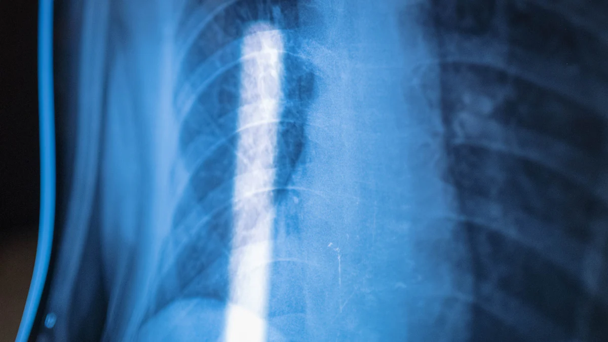
Medical imaging technologies have revolutionized diagnostics, offering precise insights into the human body. Among these, the comparison of electron beam tomography vs. CT scan reveals their unique technical capabilities. Electron beam tomography (EBT) employs a stationary electron gun and detector rings, enabling rapid imaging, particularly of moving organs like the heart. In contrast, CT scans utilize rotating X-ray beams to capture detailed cross-sectional images. While EBT excels in speed and precision, CT scans dominate in versatility and widespread application. Understanding the technical differences between these methods highlights their distinct strengths and optimal use cases.
Key Takeaways
- Electron Beam Tomography (EBT) offers ultrafast imaging, making it ideal for capturing moving organs like the heart, which enhances diagnostic accuracy.
- CT scan is versatile and widely used for diagnosing a range of conditions, providing high-resolution images that are essential for effective treatment.
- EBT excels in detecting coronary artery calcification, a key indicator of heart disease, while CT scans are better suited for general diagnostics across various body parts.
- The cost and availability of EBT systems limit their use, whereas CT scanners are more accessible and cost-effective, making them a practical choice in most healthcare settings.
- Understanding the strengths and limitations of both imaging technologies helps clinicians select the most appropriate method based on specific diagnostic needs.
Defining Electron Beam Tomography and CT Scan
Electron Beam Tomography (EBT)
Electron beam tomography, also known as electron beam computed tomography, represents a significant advancement in medical imaging. This technology employs a stationary electron gun to generate X-rays, which pass through the body to create detailed cross-sectional images. Unlike traditional CT scanners, EBT does not rely on mechanical movement. Instead, it uses stationary detector rings to capture images with remarkable speed and precision.
One of the most notable features of electron beam tomography is its ability to perform ultrafast imaging. This capability makes it particularly effective for capturing images of moving organs, such as the heart. For instance, EBT can quantify calcium deposits in coronary arteries, which helps predict the risk of coronary artery disease (CAD). Its speed and accuracy have made it a valuable tool in cardiac imaging and early disease detection.
Fact: EBT was first introduced into clinical practice in 1983 and has since been widely used for coronary artery screening.
CT Scan
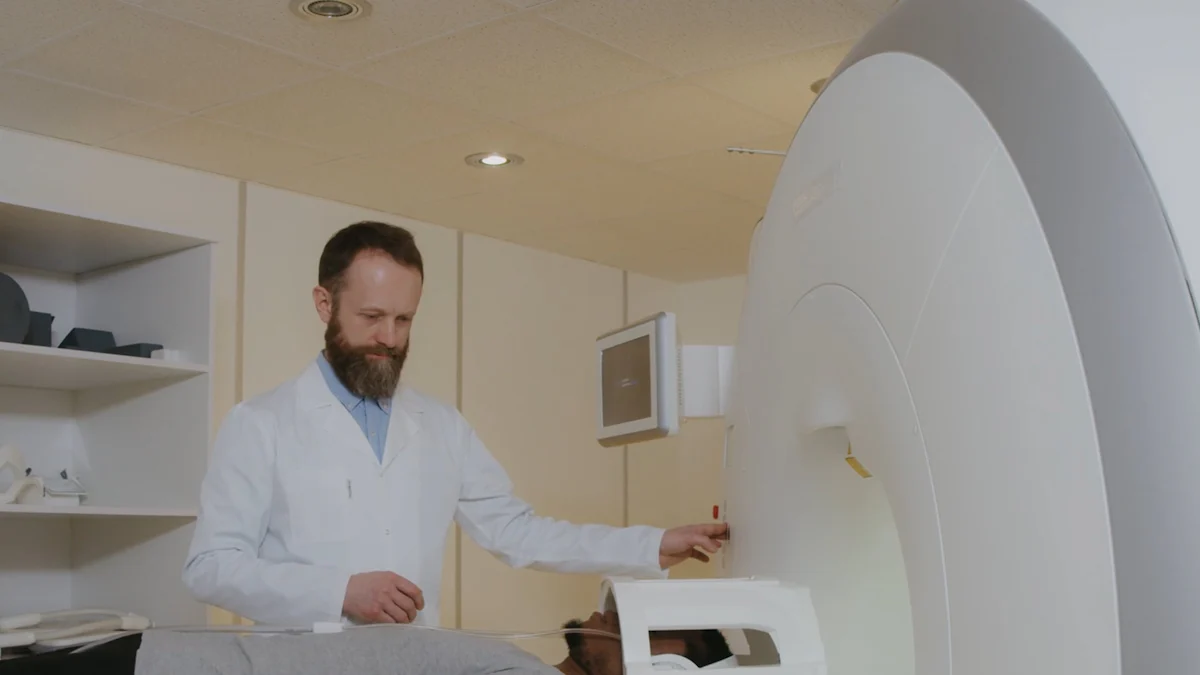
Computed tomography (CT) scan has become a cornerstone of modern diagnostic imaging. This technology uses a rotating X-ray beam to capture multiple cross-sectional images of the body. The X-ray tube and detectors rotate around the patient, taking slice-by-slice images that are later reconstructed into a detailed 3D representation.
CT scan is highly versatile and can be used to diagnose a wide range of conditions. From detecting tumors to identifying fractures, CT imaging provides high-resolution images that aid in accurate diagnosis. While not as fast as electron beam tomography, modern CT scanners, such as Ultrafast Dual Source CT scanners, have significantly improved in speed and image quality. These advancements have expanded their applications in both emergency and routine medical settings.
Did You Know? The latest CT scanners, like the Ultrafast Dual Source models, can store data from millions of scans, enhancing diagnostic accuracy through advanced data analysis.
Key Technical Differences in Electron Beam Tomography vs. CT Scan
Electron beam tomography and CT scan share the goal of creating detailed internal images, but their technical approaches differ significantly:
- Imaging Speed:
- EBT captures images almost instantaneously due to its stationary components. This makes it ideal for imaging moving organs like the heart.
- CT scans rely on rotating X-ray beams, which take slightly longer to capture each image slice.
- Technology:
- EBT uses a stationary electron gun and detector rings, eliminating the need for mechanical movement.
- CT scanners use a rotating X-ray tube and detectors, which move around the patient to capture images.
- Applications:
- EBT excels in cardiac imaging, particularly for measuring coronary artery calcium levels.
- CT scans are more versatile, suitable for diagnosing a wide range of conditions, including trauma, cancer, and vascular diseases.
- Precision and Resolution:
- EBT offers high precision for specific tasks, such as detecting coronary artery calcification.
- CT scans provide high-resolution images across various body parts, making them more adaptable for general diagnostics.
- Cost and Accessibility:
- EBT systems are less common and often more expensive due to their specialized nature.
- CT scanners are widely available and more cost-effective, making them accessible in most healthcare facilities.
Both technologies have unique strengths, and their differences highlight their complementary roles in medical imaging. Understanding these distinctions helps clinicians choose the most appropriate method based on the diagnostic requirements.
How Electron Beam Tomography and CT Scan Work
Technical Principles of Electron Beam Computed Tomography
Electron Beam Computed Tomography (EBCT) operates on a unique principle that sets it apart from traditional imaging methods. It uses a stationary electron gun to generate X-rays, which are directed toward a tungsten target. This interaction produces high-energy X-rays that pass through the body to create detailed cross-sectional images. Unlike conventional CT scanners, EBCT eliminates the need for mechanical rotation. Instead, it employs stationary detector rings to capture data, enabling rapid image acquisition.
The speed of EBCT proves crucial in imaging moving organs, particularly the heart. By capturing images in milliseconds, it minimizes motion artifacts, ensuring precise visualization of structures like coronary arteries. This capability makes EBCT highly effective for detecting coronary artery calcification, a key indicator of atherosclerosis. Studies have shown that EBCT demonstrates high sensitivity in identifying calcified plaques, correlating strongly with the presence of atherosclerotic disease.
Scientific Research Findings: Research on EBCT and Coronary Artery Calcification highlights its ability to detect calcification with remarkable accuracy. However, debates persist regarding the direct link between calcium scores and coronary events.
Technical Principles of CT Scan
Computed Tomography (CT) scan relys on a rotating X-ray tube and detectors to capture images. The X-ray tube emits a fan-shaped beam that passes through the body, while detectors measure the intensity of the transmitted rays. As the tube rotates around the patient, it collects multiple slice images, which are then reconstructed into a 3D representation using advanced algorithms.
CT technology excels in versatility. It can image various body parts, including the brain, chest, abdomen, and bones. Modern advancements, such as multislice CT (MSCT), have significantly enhanced its capabilities. MSCT uses multiple detector rows to capture several slices simultaneously, reducing scan time and improving image resolution. This innovation has expanded CT’s applications, making it indispensable in emergency settings and routine diagnostics.
Did You Know? CT scans play a pivotal role in measuring coronary artery calcium scores (CACS). This metric helps assess the risk of cardiovascular events, as noted in studies focusing on CT’s role in coronary calcification.
Comparing Imaging Mechanisms and Technologies
The imaging mechanisms of EBCT and CT scan differ fundamentally, reflecting their distinct technological approaches:
- Speed and Efficiency:
- EBCT captures images almost instantaneously due to its stationary components. This rapid imaging capability makes it ideal for dynamic organs like the heart.
- CT scans, while slower, have improved significantly with the advent of multislice technology, enabling faster and more detailed imaging.
- Image Acquisition:
- EBCT uses an electron gun and stationary detector rings, eliminating mechanical movement. This design reduces wear and tear, enhancing reliability.
- CT scanners rely on rotating X-ray tubes and detectors, which require precise synchronization to ensure accurate image reconstruction.
- Applications:
- EBCT specializes in cardiac imaging, particularly for detecting coronary artery calcification. Its precision and speed make it a valuable tool for early diagnosis of heart diseases.
- CT scans offer broader applications, ranging from trauma assessment to cancer detection. Their versatility makes them a cornerstone of modern medical imaging.
- Technological Advancements:
- EBCT remains a niche technology, primarily used in specialized cardiac imaging. Its high cost and limited availability restrict its widespread adoption.
- CT technology continues to evolve, with innovations like dual-source CT and artificial intelligence integration enhancing diagnostic accuracy and efficiency.
Scientific Insight: Electron Beam Tomography in Medical Diagnostics underscores EBCT’s role in early heart disease detection. Its speed and precision surpass traditional CT scans, particularly in cardiac imaging.
Both EBCT and CT scan contribute significantly to medical diagnostics. Their distinct mechanisms and technologies cater to different clinical needs, highlighting the importance of choosing the appropriate modality based on specific diagnostic requirements.
Advantages and Limitations of Electron Beam Tomography and CT Scan
Advantages of Electron Beam CT
Electron beam CT offers several advantages that make it a valuable tool in medical imaging. Its ability to capture images at an ultrafast speed stands out as one of its most significant benefits. This rapid imaging capability allows it to effectively visualize moving organs, such as the heart, without motion artifacts. For instance, electron beam CT excels in detecting coronary artery calcification, which serves as a critical marker for assessing the risk of coronary artery disease.
Another advantage lies in its precision. Electron beam CT provides highly accurate measurements of calcium deposits in coronary arteries. This sensitivity makes it a preferred choice for early detection of atherosclerosis. Additionally, electron beam CT exposes patients to lower levels of radiation compared to some traditional CT scans, enhancing its safety profile for repeated use in cardiac imaging.
Fact: Studies have shown that electron beam CT is more sensitive in detecting coronary artery calcification than other types of CT scans, making it a reliable tool for cardiac assessments.
Advantages of High-Resolution Imaging in CT Scan
CT scan is renowned for their high-resolution imaging capabilities, which enable detailed visualization of various body structures. This advantage makes CT scan highly versatile, allowing them to diagnose a wide range of conditions, from fractures and tumors to vascular diseases. The ability to produce clear and detailed images ensures accurate diagnoses, even in complex cases.
Modern advancements, such as multislice CT technology, have further enhanced the resolution and efficiency of CT scan. These innovations allow for simultaneous capture of multiple slices, reducing scan time while maintaining image quality. High-resolution imaging also plays a crucial role in emergency settings, where quick and accurate diagnostics are essential for effective treatment.
Did You Know? High-resolution imaging in CT scans has become indispensable in oncology, where it helps detect and monitor tumors with remarkable clarity.
Limitations of Electron Beam Tomography
Despite its advantages, electron beam tomography has certain limitations that restrict its widespread use. One major drawback is its limited availability. Electron beam CT systems are less common in healthcare facilities due to their high cost and specialized nature. This lack of accessibility can make it challenging for patients to benefit from this advanced imaging technology.
Another limitation involves its narrow scope of applications. While electron beam CT excels in cardiac imaging, it is not as versatile as traditional CT scans. Its primary focus on detecting coronary artery calcification limits its utility in diagnosing other conditions. Additionally, skepticism exists regarding the correlation between calcium scores obtained through electron beam CT and the actual likelihood of coronary events. Factors such as the presence of non-calcified plaques and the inability to pinpoint specific vulnerable lesions contribute to this uncertainty.
Scientific Insight: Research highlights that electron beam CT calcium scores may not always identify the location of severe stenosis or vulnerable plaques, which can impact its predictive accuracy for coronary events.
Limitations of CT Scan
CT scans, while versatile and widely used, come with certain limitations that impact their application and effectiveness. These drawbacks often stem from the technology’s reliance on rotating X-ray beams and its broader diagnostic scope.
- Radiation Exposure
CT scans expose patients to higher levels of radiation compared to some other imaging techniques. This increased exposure raises concerns, especially for individuals requiring repeated scans. Although advancements like low-dose CT have reduced radiation levels, the cumulative effect remains a significant consideration in long-term patient care. - Motion Artifacts
The rotating X-ray tube in CT scanners can result in motion artifacts, particularly when imaging moving organs like the heart. These artifacts may compromise image clarity and diagnostic accuracy. While modern multislice CT (MSCT) technology has improved imaging speed, it still lags behind the ultrafast capabilities of electron beam tomography (EBT). - Limited Sensitivity for Specific Conditions
CT scans may lack the sensitivity required for detecting certain conditions, such as early-stage coronary artery calcification. EBT, for instance, demonstrates higher sensitivity in identifying calcified plaques, making it more effective for specific cardiac assessments. CT scans, on the other hand, might miss subtle calcifications or non-calcified plaques, which could lead to incomplete evaluations. - Cost and Accessibility in Advanced Models
While standard CT scanners are widely available, advanced models like dual-source or ultrafast CT systems come with higher costs. These expenses can limit their accessibility in smaller healthcare facilities or regions with limited resources. Patients in such areas may not benefit from the latest technological advancements in CT imaging. - Potential Overuse in Diagnostics
The widespread availability and versatility of CT scans sometimes lead to overuse in medical diagnostics. This over-reliance can result in unnecessary radiation exposure and increased healthcare costs. Clinicians must carefully evaluate the necessity of each scan to ensure its benefits outweigh the risks.
Fact: Studies comparing EBT and CT scans highlight that ultrafast CT offers lower radiation exposure than traditional CT, but EBT still surpasses it in speed and precision for cardiac imaging.
CT scans remain indispensable in modern medicine due to their versatility and high-resolution imaging. However, understanding these limitations helps clinicians make informed decisions, ensuring the technology is used appropriately and effectively for patient care.
Comparing Electron Beam Tomography vs. CT Scan: Performance and Use Cases
Imaging Speed and Efficiency
Imaging speed plays a critical role in medical diagnostics, especially when capturing moving organs like the heart. Electron beam tomography (EBT) demonstrates exceptional high-speed imaging capabilities. Its stationary electron gun and detector rings allow it to capture images in milliseconds. This rapid imaging minimizes motion artifacts, ensuring precise visualization of dynamic structures. For instance, EBT excels in cardiac imaging by detecting coronary artery calcification with remarkable accuracy.
In contrast, CT scans rely on rotating X-ray beams to acquire images. While modern multislice CT (MSCT) technology has significantly improved imaging speed, it still lags behind EBT in terms of efficiency. CT scans require slightly more time to capture each slice, which can lead to motion artifacts when imaging fast-moving organs. However, their advancements in speed have made them suitable for emergency settings, where quick diagnostics are essential.
Reflecting on the story of Sam Rubin, who passed away from a heart attack at 63, highlights the importance of timely imaging. A coronary calcium CT scan might have detected significant calcification, potentially enabling preventive measures.
The comparison between EBT and traditional CT scans reveals that EBT’s speed makes it ideal for specialized applications like cardiac imaging. CT scans, while slower, remain versatile and effective for broader diagnostic purposes.
Image Quality and Resolution
The resolution of medical imaging technologies determines their ability to produce detailed images. EBT offers superior image resolution for specific tasks, such as detecting calcified plaques in coronary arteries. Its stationary design eliminates mechanical movement, reducing the risk of image distortion. This precision enhances diagnostic accuracy, particularly in identifying early signs of atherosclerosis.
CT scans, on the other hand, provide high-resolution imaging across various body parts. Their ability to produce detailed images makes them indispensable in diagnosing conditions like fractures, tumors, and vascular diseases. Modern CT scanners, equipped with multislice technology, capture multiple slices simultaneously. This innovation improves resolution and reduces scan time, ensuring clear and accurate diagnostics.
Did You Know? High-resolution imaging in CT scans has become a cornerstone in oncology, aiding in the detection and monitoring of tumors with exceptional clarity.
While EBT specializes in cardiac imaging, CT scans offer enhanced diagnostic accuracy for a wide range of conditions. Both technologies contribute significantly to medical diagnostics, with their resolution tailored to different clinical needs.
Cost and Accessibility
Cost and accessibility often influence the choice between electron beam tomography vs. CT scan. EBT systems are less common due to their high cost and specialized nature. Their limited availability restricts their use to specific healthcare facilities, primarily those focusing on cardiac imaging. This exclusivity makes EBT less accessible to patients in regions with limited resources.
CT scans, in contrast, are widely available and more cost-effective. Standard CT scanners are present in most healthcare facilities, making them accessible to a larger population. Advanced models, such as dual-source CT scanners, come with higher costs but offer improved capabilities. Despite their expense, these models remain more affordable and accessible than EBT systems.
An assessment by the National Coordinating Centre for Health Technology Assessment highlighted the cost-effectiveness of CT scans. While CT screening can detect arterial calcification, its affordability and versatility make it a practical choice for routine diagnostics.
The comparison between EBT and CT scans underscores their complementary roles. EBT’s high-speed imaging and precision cater to specialized applications, while CT scans provide a cost-effective solution for general diagnostics. Understanding these differences helps clinicians choose the most appropriate technology based on patient needs and healthcare resources.

Conclusion
Electron Beam Tomography (EBT) and CT scan showcase distinct technical strengths, catering to different diagnostic needs. EBT excels in speed and precision, particularly for cardiac imaging, due to its stationary electron beam technology. CT scans, with their rotating X-ray systems, offer versatility and high-resolution imaging across various medical applications.
Both technologies play pivotal roles in advancing diagnostics and research. EBT’s rapid imaging aids in early detection of heart conditions, while CT scans remain indispensable for broader diagnostic purposes. Future advancements in these imaging methods promise enhanced accuracy, reduced radiation exposure, and expanded accessibility, further revolutionizing medical diagnostics.